- What causes a fetlock fracture in a horse?
- What is the function of the fetlock?
- What is the fetlock joint in a horse?
- How does a sesamoid fracture affect a horse?
- What is the lameness of the fetlock joint in horses?
- What is the pastern of a horse?
- What is a horse’s first phalanx?
- What is at the rear of the fetlock joint?
- What is a horse fetlock?
- What is a fetlock injury?
- Why is the fetlock joint so easy to image accurately?
- What is the fetlock joint?
- What causes sesamoid fractures in Standardbreds?
- How do you diagnose sesamoid injuries in horses?
- What are fractures of the proximal sesamoid bones?
- Where is the proximal sesamoid bone in a horse?
- What causes a horse’s legs to fill up with fluid?
- What causes osselets in horses to cause pain?
- What kind of injury can a horse get from its elbow?
- What is the pastern bone on a horse?
- What determines the length of a horse’s pastern joint?
- What is a P1 fracture in a horse?
What causes a fetlock fracture in a horse?
They usually occur when the horse is exercising at a high speed due to overextension (hyperextension) of the fetlock joint. An affected horse will suddenly become lame and have swelling of the fetlock joint. Another type of fracture involves chips or loose fragments on the back of the long pastern bone.
What is the function of the fetlock?
The fetlock is a joint, a shock absorber, an energy storage system and a stabilizer of the front limbs. It is a complex joint with the bones and soft tissue interwoven.
What is the fetlock joint in a horse?
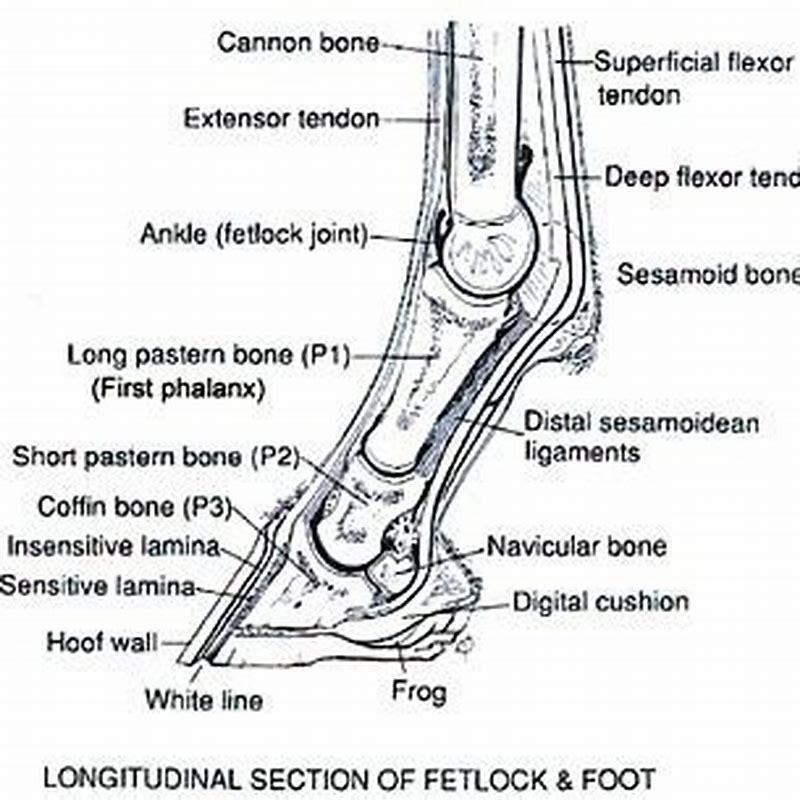
Fetlock Joint – The fetlock joint occurs between the cannon bone, the proximal phalanx and sesamoid bones in the front legs. It allows bending and extension movements. The fetlock joint is arguably the joint that distinguishes a horse, with its unique anatomy and physiology allowing high speed, medium distance activity.
How does a sesamoid fracture affect a horse?
A horse will usually suffer from immediately lameness upon fracturing the sesamoid. The fetlock joint will frequently be pulled downward because of lack of support following the fracture. The lowered fetlock causes more weight and pressure on the heel, which forces the toe to point up. The fetlock may also be hyperextended.
What is the lameness of the fetlock joint in horses?
Lameness of the fetlock joint in horses is fairly common. The fetlock joint (metacarpophalangeal/metatarsophalangeal joint) is a high motion joint with a small cross-sectional area and little soft tissue covering, which makes it predisposed to injury.
What is the pastern of a horse?
The pastern is the area between the hoof and the fetlock joint. Disorders of the fetlock and pastern include conditions such as fractures, osteoarthritis, osselets, ringbone, sesamoiditis, synovitis, and windgalls. Horses’ legs are complex and easily injured.
What is a horse’s first phalanx?
Horses’ legs are complex and easily injured. Fractures of the long pastern bone (first phalanx) are not uncommon in racehorses or other performance horses. They may be small “chip” fractures, fractures along the length of the bone (split pastern), or comminuted fractures in which the bone is broken into multiple fragments.
What is at the rear of the fetlock joint?
At the rear of the fetlock joint is a small bone called the sesamoid. Unlike humans ankles, the horse’s leg has no muscles and are in fact more similar to our fingers than our arms or legs.
What is a horse fetlock?
A ‘horses fetlock’ is a name of a joint between the horses cannon bone and pastern bone and is ‘the ankle’ of a horse. At the rear of the fetlock joint is a small bone called the sesamoid.
What is a fetlock injury?
The fetlock joint can show inflammation (referred to as synovitis or capsulitis) and the supporting ligaments (Suspensory ligament branch and the Distal Sesamoidean Ligaments) can also become inflamed (Sesamoiditis). Injuries to the sesamoid bones (the back of the fetlock) are now being recognised as a common fetlock injury in jumping horses.
Why is the fetlock joint so easy to image accurately?
Dean W. Richardson, Sue J. Dyson, in Diagnosis and Management of Lameness in the Horse (Second Edition), 2011 The fetlock joint is easy to image accurately because of the size and accessibility.
What is the fetlock joint?
Fetlock is a term used for the joint where the cannon bone, the proximal sesamoid bones, and the first phalanx (long pastern bone) meet. The pastern is the area between the hoof and the fetlock joint.
What causes sesamoid fractures in Standardbreds?
The lateral proximal sesamoid in the hindlimb of Standardbreds may be fractured as a result of torque forces induced by shoeing with a trailer-type shoe. Other fractures include mid-body, basilar, abaxial, axial, or comminuted, and they may involve one or both sesamoids.
How do you diagnose sesamoid injuries in horses?
High speeds coupled with suspensory apparatus anatomy can lead to sesamoid injuries in horses. Fracture diagnosis can be tricky because changes aren’t always evident when using traditional methods such as palpation and radiographs. Veterinarians have determined that nuclear scintigraphy, MRI, and CT are good diagnostic tools to detect problems.
What are fractures of the proximal sesamoid bones?
What are Fractures of the Proximal Sesamoid Bones? Fractures of the proximal sesamoid bones can happen to any horse but they are more common in racing Thoroughbreds, Standardbreds, and Quarter horses. Fractures to the proximal sesamoid bones are classified by where the fracture is located.
Where is the proximal sesamoid bone in a horse?
Proximal sesamoid bones are located in the lower limbs of the horse and are part of the metacarpophalangeal (fetlock) hinge joint. The hyperextension of the metacarpophalangeal joint causes extreme tension across the proximal sesamoid bones, which can cause them to fracture. Symptoms of Fractures of the Proximal Sesamoid Bones in Horses
What causes a horse’s legs to fill up with fluid?
Filled legs of this nature are caused by an abnormal accumulation of fluid in the tissues called oedema and will go down with gentle exercise or when the horse is turned out. Also Know, how do you reduce swelling in a horse’s knee? Unfortunately, if damaged, carpal tendon sheaths tend to produce excess synovial fluid that results in a ‘big knee’.
What causes osselets in horses to cause pain?
The condition is an occupational hazard for young Thoroughbreds and is caused by the strain and repeated trauma of hard training in young horses. The gait of a horse with osselets becomes short and choppy. Applying firm pressure and bending the fetlock joint will cause pain.
What kind of injury can a horse get from its elbow?
Collateral Ligament Injury in the Elbow of Horses Disorders of the Metatarsus in Horses Bucked Shins/Dorsal Cortical Fractures of the Third Metatarsal Bone in Horses Exostoses of the Metatarsal Bones in Horses
What is the pastern bone on a horse?
(April 2008) The pastern is a part of the leg of a horse between the fetlock and the top of the hoof. It incorporates the long pastern bone (proximal phalanx) and the short pastern bone (middle phalanx), which are held together by two sets of paired ligaments to form the pastern joint (proximal interphalangeal joint).
What determines the length of a horse’s pastern joint?
Conformation. The length of the pastern joint is determined by the length of the first phalanx. The short pastern bone is less a determinant because it is smaller, at 2 inches in length, and part of it is encased in the hoof.
What is a P1 fracture in a horse?
Fractures of the first/proximal phalanx (P1) may occur in any type of horse used for performance. They may be small osteochondral “chip” fractures along the dorsal margin of the proximal joint surface, sagittal (complete or incomplete), or comminuted.