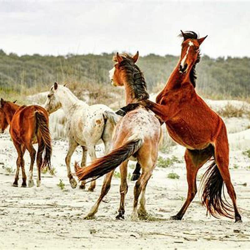
- How to tell if a horse has a gut infection?
- Can ultrasonography be used to assess gastrointestinal motility in horses?
- Is ultrasound superior to rectal examination for diagnosing intestinal obstruction in horses?
- What is intestinal volvulus in a horse?
- What is abdominal ultrasonography for horses with colic?
- What is ultrasonography used for in horses?
- How do you label an ultrasonographic image of a horse?
- Why is ultrasound not used to examine the bone?
- How does ultrasound work?
- How do you diagnose intestinal obstructions in horses?
- What is abdominal ultrasonography for acute colic?
- How to diagnose colic in horses with abdominal pain?
- How many horses have been referred for colic in 2 months?
- What causes Si volvulus in horses?
- What is the pathogenesis of volvulus?
- Can you do ultrasonography without clipping a horse?
- Which transducer is best for imaging suspensory ligament in horses?
- Can we measure the conformation of jumping thoroughbred horses?
- Is a bone scan a valid diagnostic screening test for Sport Horses?
- Is Nuclear scintigraphy a good imaging method for horses?
- How does ultrasound work in veterinary medicine?
- Can ultrasonic sound be used to diagnose and treat horses?
- What tests are used to diagnose a horse’s disease?
- What causes dilatation of the intestine in horses?
How to tell if a horse has a gut infection?
Using a stethoscope is the best way to hear gut sounds. Put the stethoscope up against the horse’s barrel just behind his last rib. If you hear lots of sounds, the horse is probably all right. Be sure to check from both sides. HorseCoursesOnline.com
Can ultrasonography be used to assess gastrointestinal motility in horses?
1 Department of Clinical Studies, New Bolton Center, University of Pennsylvania, School of Veterinary Medicine, Kennett Square, Pennsylvania, USA. Background: Auscultation and ultrasonography are noninvasive techniques used to assess gastrointestinal motility in horses.
Is ultrasound superior to rectal examination for diagnosing intestinal obstruction in horses?
Likewise, the diagnosis of small intestinal obstruction was made accurately after sonographic examination in all affected horses in the present study. Therefore, the authors feel that ultrasound is superior to rectal examination for this diagnosis.
What is intestinal volvulus in a horse?
Ultrasonographic image of a horse with an intestinal volvulus (large amount of hemorrhagic abdominal fluid), ven- tral abdomen. {1) Dilated loop of small intestine; (2) large amount of hemorrhagic abdominal fluid. Reproduced with kind permission from Scharner and co-workers Pferde- heilkunde 18:57-63, 2002.
What is abdominal ultrasonography for horses with colic?
Abdominal ultrasonography has been part of the examina- tion protocol of each horse with acute colic admitted to the Surgical Clinic of the Veterinary Medical Faculty of the Univer- TABLE 1. UItrasonography of the Equine Acute Abdomen Name of Window Organs 1. Ventral abdomen (xyphoid cartilage to inguinal area) 2. Stomach window 3.
What is ultrasonography used for in horses?
Ultrasonography has proven to be very useful for evaluation of the urinary and gastrointestinal tracts in foals and adult horses.
How do you label an ultrasonographic image of a horse?
All ultrasonographic images should be labeled with the date of examination, the horse’s name, the owner’s or trainer’s name, the limb being examined, and the location of the image. In addition, it is helpful to include the age, breed, and use of the horse and the current exercise level.
Why is ultrasound not used to examine the bone?
• Ultrasound cannot be used to examine the deeper structures in bone as bone matter is too dense • Problems can arise when ultrasound is used to look at curved tendons and ligaments because if the surface is not parallel to the ultrasound, the sonic beam will be reflected elsewhere How much does ultrasound cost?
How does ultrasound work?
“Sound waves leave the transducer and enter the body, where they reflect off the bone and soft tissue to produce an echo that is analysed by a computer in the ultrasound machine and transformed into moving pictures of the organ or tissue that is being examined.
How do you diagnose intestinal obstructions in horses?
Intestinal obstructions are diagnosed based on the horse’s history, signs, and physical and rectal examination findings. Ultrasonography and analysis of abdominal fluid may also help make the diagnosis. Some obstructions, however, require surgery to make a definitive diagnosis.
What is abdominal ultrasonography for acute colic?
Abdominal ultrasonography has been part of the examina- tion protocol of each horse with acute colic admitted to the Surgical Clinic of the Veterinary Medical Faculty of the Univer- TABLE 1. UItrasonography of the Equine Acute Abdomen Name of Window Organs 1.
How to diagnose colic in horses with abdominal pain?
More recently, abdominal radiography and abdominal ultrasonography have become more useful in evaluating horses with abdominal pain. Several recent studies have shown that abdominal ultrasonog- raphy in the colic patient can be a very useful diagnos- tic tool. Technique Abdominal ultrasonography is a very safe and non- invasive diagnostic test.
How many horses have been referred for colic in 2 months?
The clinical subjects comprised 36 horses that had been referred for colic over a 2 month period. Each horse was examined at admission and FLASH findings at seven topographical locations were compared to serial clinical examinations, surgical and non-surgical outcomes, or with post-mortem reports.
What causes Si volvulus in horses?
A small intestinal (SI) volvulus occurs in the horse when the intestine rotates on its mesenteric axis through an angle greater than 180o, resulting in strangulation. The pathogenesis is thought to be due to a segment of hypermotile intestine preceding a segment where peristalsis has ceased, leading to the development of a twist.
What is the pathogenesis of volvulus?
The pathogenesis is thought to be due to a segment of hypermotile intestine preceding a segment where peristalsis has ceased, leading to the development of a twist. Volvulus may occur as a primary cause of colic or secondarily as a result of another condition such as a lipoma, incarceration within the mesentery or adhesion within the abdomen.
Can you do ultrasonography without clipping a horse?
As regards the hind limb, clipping should involve the metatarsal region up to the chestnut. It is possible in some thin-haired horses to perform ultrasonography without clipping, but the image quality will always be impaired and an acceptable image may not be obtained in all cases.
Which transducer is best for imaging suspensory ligament in horses?
High-frequency (7.5–16 MHz) linear array transducers are preferred. A micro-convex transducer may be useful to image the proximal aspect of the suspensory ligament in some horses.
Can we measure the conformation of jumping thoroughbred horses?
Therefore the aim of the present study was to use objective methods to establish a set of baseline measurements of conformation in jumping thoroughbred horses. This study was carried out on 51 jumping thoroughbred horses belonging to Egyptian Armed Forces Equestrian Club. These horses were of different ages (5-15 years).
Is a bone scan a valid diagnostic screening test for Sport Horses?
But researchers hadn’t verified its validity as a diagnostic screening test in lame or poorly performing sport horses. So, Dyson and colleagues reviewed 690 bone scans performed at the Centre for Equine Studies from March 2008 to December 2014.
Is Nuclear scintigraphy a good imaging method for horses?
In theory, nuclear scintigraphy (bone scanning) sounds like the ideal imaging modality to localize problem areas in horses’ bodies. Veterinarians inject a radioisotope into the horse, which gets deposited in areas where bone is changing rapidly, as often occurs at healing injury sites.
How does ultrasound work in veterinary medicine?
What is ultrasound and how does it work? Roger Smith MRCVS, professor of equine orthopaedics at the Royal Veterinary College, explains: “Ultrasound imaging uses the principles of sonar developed for ships at sea. “As sound passes through the body, it produces echoes that can identify the distance, size and shape of objects.
Can ultrasonic sound be used to diagnose and treat horses?
The use of ultrasonic sound for diagnosis and treatment in human and equine medicine is not new, and in fact is becoming commonplace. Most horse breeders, for example, are familiar with the use of diagnostic ultrasound to detect and monitor
What tests are used to diagnose a horse’s disease?
Other tests can include blood counts and cultures, electrolyte analysis, and joint, cerebrospinal or tracheal fluid testing in foals.
What causes dilatation of the intestine in horses?
Generally, the lumen of the intestine is completely obstructed, such as occurs with a strangulating obstruction, enterolithiasis Enterolithiasis The most common cause of gastric dilatation in horses is excessive gas or intestinal obstruction.