- What age do horses knees close?
- Is a 4 year old horse a full grown horse?
- When do horses knees close up?
- When should you X-ray your horse’s knee?
- Can you ride a young horse with open knees?
- Why do horses have bones in their knees?
- How many bones are in a horse’s knee?
- When does a horse’s hip bone stop growing?
- What does it mean when a horses knees close?
- Do X-rays show arthritis in horses?
- Is it safe to sedate a horse for X rays?
- Why do they take Xrays of horses in the stable?
- How many X-rays do I need when buying a horse?
- Are your horses’ knees prone to congenital and acquired problems?
- Why do horses knees knock together when they walk?
- Are your horse’s knees prone to lameness?
- What is the last bone to fuse in a horse?
- How do X-rays work on horses?
- How do you know if your horse has joint problems?
- Can a horse with osteoarthritis show up on a Xray?
- Can a horse have a normal X ray and still be lame?
- What are the different types of X-rays used to treat horses?
- Can a horse be scanned under standing sedation?
- Can X-rays detect soft-tissue injuries in horses?
What age do horses knees close?
However, they typically reach their maximum height at four or five years of age. The bones of horses have cartilage on either end of each bone in their body, and as the horse ages, the bones fuse, creating a bond. The bones are used to determine the stages of horse growth. A horses’ knees commonly close around two years old.
Is a 4 year old horse a full grown horse?
Four-year-old horses are considered adults. Once a horse reaches four, they are considered adult horses. Between four years old and five years old, a horse will stop growing any taller. As a horse reaches six years old, he is likely fully grown, the only way to confirm this is with an x-ray.
When do horses knees close up?
A horses’ knees commonly close around two years old. Owners and trainers usually check a horse to make sure their knees are fused before breaking him to ride.
When should you X-ray your horse’s knee?
If you think you can determine this by feeling the horse’s knee, think again. It’s tough to see any of this from the outside, said Ross Cleland, DVM, Carlton, Ore. He advises you to x-ray the knees to see how the closing is progressing around 24 to 30 months of age. Larger breeds may continue into their three-year-old year.
Can you ride a young horse with open knees?
A young horse fed an inadequate or unbalanced diet is likely to experience even more problems if you start riding him with open knees, especially if you ride him hard. The most common problem you may encounter is the creation of an inflammation in the growth plate.
Why do horses have bones in their knees?
The bones of horses have cartilage on either end of each bone in their body, and as the horse ages, the bones fuse, creating a bond. The bones are used to determine the stages of horse growth. A horses’ knees commonly close around two years old.
How many bones are in a horse’s knee?
Horses’ knees, which are the equivalent to our wrists, are each made up of two rows of bones that flex in three different places—though markedly less in the bottom joint. Add to that stack of bones an extensive network of tendons and ligaments, and you have a sophisticated structure that’s
When does a horse’s hip bone stop growing?
Pelvis – the hip socket is firm between 18mos. and 2 years, but the rest of the bone does not stop growing until the horse is 5 or more years old. And what do you think is last? The vertebral column (spine) of course.
What does it mean when a horses knees close?
The process a young horse goes through as his bones develop and his knees “close” actually involves the growth plate, not the knee joint. The growth plates, which are above and below the joint, are modified forms of cartilage that calcify and lengthen the bone.
Do X-rays show arthritis in horses?
Otherwise stated, people with some rather ugly X-rays of their knee can show very little in the way of clinical signs of arthritis, especially pain, and people who are in a lot of pain can have X-rays that don’t look very bad at all. I suspect it’s the same way in horses.
Is it safe to sedate a horse for X rays?
Because X-rays pose a health risk to humans and animals, it is important that the horse is correctly restrained so that there is no ‘scatter’. Proper positioning is also essential to prevent misdiagnosis. This may require sedation or even full anaesthesia.
Why do they take Xrays of horses in the stable?
They are used to examine the anatomy of the internal structures of horses, either for diagnosis of a disease or before purchasing a horse to confirm that it is sound. Most equine practices have a portable X-ray machine that can be used for examining the horse in the stable.
How many X-rays do I need when buying a horse?
A routine pre-purchase ‘protocol’ includes three sets of X-rays of the foot and navicular bone for each leg (12 images); front to back and side views of each fetlock (8 images); front to back, side views and two oblique views of each hock (8 images). In addition, radiographs of the stifle joints are advised in young horses.
Are your horses’ knees prone to congenital and acquired problems?
Horses’ knees are prone to both congenital and acquired lameness problems. Here’s what you need to know. U p to 90% of lamenesses affecting horses’ front legs stem from bone and soft tissue found from the fetlock joint down.
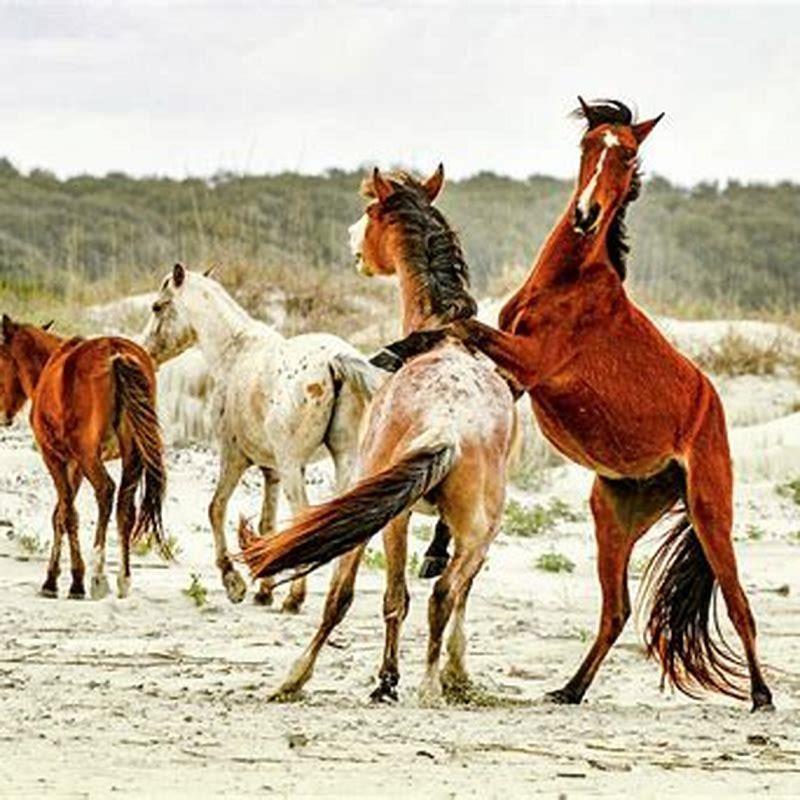
Why do horses knees knock together when they walk?
Your horse may have a distinctive gait where the knees knock together as they walk or run. This condition is caused due to the uneven growth of the leg bones. As the bones grow unevenly the muscles and other bones may attempt to compensate for this uneven growth, causing further deviations and contortion.
Are your horse’s knees prone to lameness?
Horses’ knees are prone to both congenital and acquired lameness problems. Here’s what you need to know. It’s now widely known that preexisting conditions in racehorses can be a telltale sign of breakdown risk. U p to 90% of lamenesses affecting horses’ front legs stem from bone and soft tissue found from the fetlock joint down.
What is the last bone to fuse in a horse?
Femur – there are 4 major epiphyses on this bone, including the head that goes into the hip socket; they fuse between 3 – 4 years. Pelvis – the hip socket is firm between 18mos. and 2 years, but the rest of the bone does not stop growing until the horse is 5 or more years old. And what do you think is last? The vertebral column (spine) of course.
How do X-rays work on horses?
And just like visible light rays, X-rays do one of two things. Either they pass through stuff, or stuff gets in the way of them (in a variety of ways, such as absorption, or reflection, which we’re not going to talk about right now). Pretty simple, actually. So, when you shoot X-rays at a horse, mostly, one of three things can happen:
How do you know if your horse has joint problems?
To check your horse’s muscle and joint functions, the veterinarian will examine the way the muscles work and manipulate the joints to check for restricted movements. Radiographs (x-rays) are essential to diagnosing your horse and will show inflammation, thickening of the tissues around the cartilage, and decreased space in the joints.
Can a horse with osteoarthritis show up on a Xray?
Radiographs (X rays) reveal typical OA lesions, but none might be evident in the early stages because cartilage is not visible on radiographs. Horses with mild OA often appear stiff when emerging from the stall or starting work before warming up and looking more comfortable.
Can a horse have a normal X ray and still be lame?
Not all horses with abnormal X rays will go on to develop lameness. Conversely, a horse which shows lameness during the examination may have normal X rays.” Powell explains that what you might expect to find in an apparently sound horse’s foot varies, depending on age and how many miles are on the clock.
What are the different types of X-rays used to treat horses?
• Magnetic resonance imaging (MRI). • Computed tomography (CT). Radiography is the oldest and still the most commonly used imaging modality in equine practice. How are X-rays produced?
Can a horse be scanned under standing sedation?
This development has led to CT becoming more accessible for equine veterinary use, although the scanner at Rossdales is one of only 7 systems in the UK capable of scanning horses under standing sedation. Standing CT in adult horses is limited to the head, neck and limbs; the amount of each depending on the individual system.
Can X-rays detect soft-tissue injuries in horses?
Again, veterinarians are unlikely to note soft-tissue injuries via radiography, but they can use X rays to rule out bone and/or joint involvement in injured horses, such as those with wounds or blunt trauma from a horse kicking a stall wall, for instance.