- How many colors are used in FISH analysis?
- How to make your fish more colorful?
- What is fish used to diagnose?
- What are Multicolor FISH assays?
- What determines the color produced by a chromatophore?
- What are the types of chromatophores in fish?
- How to make fish’s color better?
- Do fish get more colorful as they age?
- How can I improve my fish’s coloration?
- Why is my male fish getting more colorful?
- What is the function of fish in Chromosome Biology?
- How is fish used to diagnose genetic disorders?
- What are the components of a fish assay?
- Can Multicolor-FISH assays be used for chromosomal differentiation?
- What is multicolor FISH used for?
- What are sky and M-FISH assays?
- What diseases can be diagnosed using fish?
- What is the FISH technique in genetic testing?
- What is the difference between fishing tackle and fishing techniques?
- What is the FISH technique?
- What is m-fish used for?
- What are the three types of pigment cells?
- Are pigment cells common in fish?
How many colors are used in FISH analysis?
Traditional FISH analysis has employed, at most, two colors of detection, a red-fluorescing fluorochrome and a green-fluorescing fluorochrome. The improvements in fluorescent imaging and development of heartier fluorochromes/dyes have enabled investigators to use several different DNA probes in one …
How to make your fish more colorful?
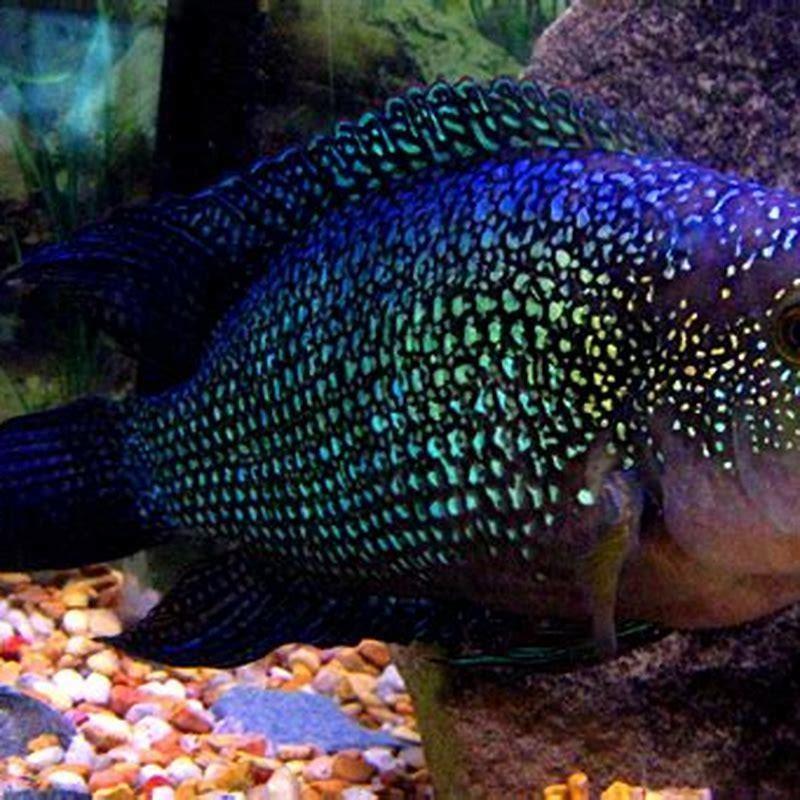
You can make your fish more colorful by feeding them color-enhancing foods such as brine shrimp, scallops, Mysis shrimp, romaine lettuce, broccoli, and spinach. You should now have plenty of information to help you make your decision about which colorful fish you want to fill your tank with.
What is fish used to diagnose?
In medicine, FISH can be used to form a diagnosis, to evaluate prognosis, or to evaluate remission of a disease, such as cancer. Treatment can then be specifically tailored.
What are Multicolor FISH assays?
Multicolor FISH (M-FISH) assays are used for a precise assessment of complex chromosomal rearrangements. This technique uses all whole-chromosome painting probes in multiplex-FISH and spectral karyotyping.
What determines the color produced by a chromatophore?
The color produced by the unit depends largely upon the color of pigment in the xanthophore and the reflectivity of the iridophore. Melanophores are largely responsible for lightening or darkening of the color produced in the other two chromatophores.
What are the types of chromatophores in fish?
Seven distinct sorts of chromatophore can be found in fish— iridophores, leucophores, melanophores, xanthophores, erythrophores, cyanophores anderythro-iridophores (Hickman et al, 1995; Goda et al, 2013). Iridophores contain schemochromes.
How to make fish’s color better?
Keeping the fish tank water conditions ideal for your fish will enhance colors naturally. Some fish food can enhance fish colors. Good lighting will help to enhance your fish’s colors as well. Below we will explore these options so you too can have a vibrant, healthy tank with bright, beautiful fish. Why Worry About the Color of Fish?
Do fish get more colorful as they age?
As a result, some fish species will become more colorful as they age. Some fish colors will become brighter when they are in the mood for spawning. Both male and female of the same species might have the brightest colors during mating seasons.
How can I improve my fish’s coloration?
Decorating your tank to imitate the natural habitat of your fish can have a significant impact on improving their coloration. Some fish prefer dark substrate over light substrate, for example, based on their natural habitat.
Why is my male fish getting more colorful?
During mating seasons, for example, male fish may develop more intense coloration to attract a mate and females may become more colorful as they begin to produce eggs. If you plan to breed your fish, feeding them a healthy diet will not only encourage healthy coloration but it will also encourage breeding behavior.
What is the function of fish in Chromosome Biology?
It is used to detect and localise the presence or absence of specific DNA sequences on chromosome. FISH involves hybridising a fluorescent labelled DNA probe to denatured chromosomal DNA of metaphase chromosome, as well as I phase chromosomes.
How is fish used to diagnose genetic disorders?
From a medical perspective, FISH can be applied to detect genetic abnormalities such as characteristic gene fusions, aneuploidy, loss of a chromosomal region or a whole chromosome or to monitor the progression of an aberration serving as a technique that can help in both the diagnosis of a genetic disease or suggesting prognostic outcomes.
What are the components of a fish assay?
There are two major elements required in a conventional FISH assay: the probe and the target sequence. In the very first step, before doing any wet lab work we have to select the sequence or the portion of a chromosome we wish to study. The FISH is capable of detecting the fragments of DNA of more than a few thousand of base pairs.
Can Multicolor-FISH assays be used for chromosomal differentiation?
Since that time different approaches for chromosomal differentiation based on multicolor-FISH (mFISH) assays have been published with the purpose to characterize structurally abnormal chromosomes and supernumerary marker chromosomes of unknown origin after conventional karyotypic analysis.
What is multicolor FISH used for?
Multicolor FISH. Multicolor-FISH (mFISH) is a method to facilitate analysis of each single chromosome or chromosome part of a metaphase. Thus, marker chromosomes, complex chromosomal rearrangements, and all numerical aberrations can be visualized simultaneously in a single hybridization experiment.
What are sky and M-FISH assays?
The generic term for multi-color FISH assays is M-FISH, however, the technologies behind the manner in which the fluorochrome information is generated has spawned two different M-FISH systems: spectral karyotyping (SKY) and M-FISH.
What diseases can be diagnosed using fish?
The diseases that have been diagnosed using FISH include Prader-Willi syndrome, Angelman syndrome, 22q13 deletion syndrome, chronic myelogenous leukemia, acute lymphoblastic leukemia, Cri-du-Chat syndrome, velocardiofacial syndrome, and Down syndrome. The analysis of chromosomes 21, X, and Y can identify oligozoospermic individuals at risk.
What is the FISH technique in genetic testing?
The technique relies on exposing chromosomes to a small DNA sequence called a probe that has a fluorescent molecule attached to it. FISH helps scientist to visualize the location of particular gene to check for a variety of chromosomal abnormalities.
What is the difference between fishing tackle and fishing techniques?
Fishing tackle refers to the physical equipment that is used when fishing, whereas fishing techniques refers to the manner in which the tackle is used when fishing. It is possible to harvest many sea foods with minimal equipment by using the hands.
What is the FISH technique?
The FISH technique is dependent upon hybridizing a probe with a fluorescent tag, complementary in sequence, to a short section of DNA on a target gene. The tag and probe are applied to a sample of interest under conditions that allow for the probe to attach itself to the complementary sequence in the specimen if it is present.
What is m-fish used for?
The M-FISH is known as multicolor FISH uses different colored probes for different chromosomes. Broadly, it is used in the characterization of different chromosomes and numerical chromosomal abnormalities. Known as quantitative FISH used for the quantification of the genetic material hybridized by the probe.
What are the three types of pigment cells?
The three basic pigment cell types found in poikilothermic vertebrates, melanocytes (melanin-producing cells), erythrophores (red or yellow pigment cells), and iridophores (iridescence-producing cells), are derived from neural crest.
Are pigment cells common in fish?
These pigment cell tumors are among the most common types in bony fish and seem to be more common in fish than in mammals, including humans. Moreover, there are no mammalian neoplasms corresponding to erythrophoromas and iridophoromas in fish.


