- Can you get a charley horse at any time?
- Can horses have straight hind limbs?
- How to pick up a horse’s back legs?
- How much fluid in the carpal sheath of a horse?
- How far does the carpal synovial sheath extend?
- What is the anatomy of the carpal canal?
- How to flex a horse’s shoulder?
- What is a synovial sheath?
- What is the difference between synovial sheath and paratenon?
- What is equine carpal joint disease?
- What is the structure of the carpal joint?
- How to diagnose tendon sheath injuries in horses?
- How do you assess fluid on a horse’s leg?
- How to tell if a horse has swelling in the carpus?
- What are synovial bursae and tendon sheaths?
- What is the function of the synovial fluid?
- What is the function of the synovial sheath?
- What is the anatomy of the paratenon?
- How are the flexor tendons attached to the fibrous flexor sheath?
- Where is the accessory carpal bone located?
- What are the symptoms of carpal hygroma in horses?
- What does it mean when a horses carpus is swollen?
- How do you know if your horse has carpal tunnel syndrome?
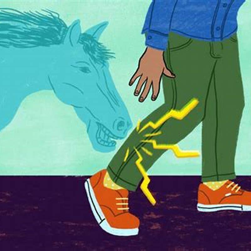
Can you get a charley horse at any time?
You can get a charley horse at any time, but up to 60% of adults experience them at night. They can also strike during athletic activities that involve kicking motions, such as running and swimming.
Can horses have straight hind limbs?
Nearly any breed can have straight hind limbs, which predispose horses to suspensory ligament (which extends down the back of the lower leg between the cannon bone and the deep digital flexor tendon) injuries or degenerative conditions from repeated overloading.
How to pick up a horse’s back legs?
Always let the horse know you are about to pick up their back legs by running your hand alongside them and running your hand down the leg to pick it up especially if you are not familiar with the horse. Hold each stretch for as long as the horse will allow you but ideally 30 seconds is an adequate stretch and repeat 3 times on each leg.
How much fluid in the carpal sheath of a horse?
In clinically normal horses, the amount of fluid within the carpal sheath varies, but it is usually the same bilaterally in each horse.
How far does the carpal synovial sheath extend?
The carpal synovial sheath extends from 7 to 10 cm proximal to the antebrachiocarpal joint to the midmetacarpal region. The proximal recess is wide and extends between the ulnaris lateralis and lateral digital extensor muscles laterally, but it is firmly supported on the medial aspect by the antebrachial fascia.
What is the anatomy of the carpal canal?
Anatomy. The carpal canal encloses the carpal synovial sheath, which contains the superficial (SDFT) and deep (DDFT) digital flexor tendons. The dorsal wall of the carpal canal is formed by the common palmar ligament of the carpus, which is a thickened part of the fibrous joint capsule that extends distally as the accessory ligament of the DDFT…
How to flex a horse’s shoulder?
To flex the shoulder, pick up the hoof and hold the leg at a 90-degree angle for 10 to 15 seconds before putting the foot back down.
What is a synovial sheath?
Synovial Sheath. A synovial sheath is a sac that completely surrounds a tendon, forming a synovial lining on the surface of the tendon and the lining of the sheath.
What is the difference between synovial sheath and paratenon?
When not within a synovial sheath, the tendons are surrounded by the paratenon, a layer of connective tissue that overlies loose connective and vascular tissue comprising the epitenon. The flexor tendons are enclosed by synovial sheaths. In the fibrous flexor sheath both tendons are invested by a common synovial sheath.
What is equine carpal joint disease?
Equine carpal joint disease is seen most commonly as an ‘occupational disease’ of racehorses and occurs less commonly in horses used for other purposes. A form of traumatic arthritis/osteoarthritis (OA) appears to be the usual underlying disease entity.
What is the structure of the carpal joint?
The carpal joint consists of two rows of bones and three joint levels, the antebrachiocarpal, the middle carpal, and the carpometacarpal joints. The individual bones are connected to the joint capsule and there are numerous short ligaments, most of them only spanning one joint level.
How to diagnose tendon sheath injuries in horses?
“Diagnostic techniques most frequently used to identify tendon sheath injury or damage include ultrasonography and tenoscopy, which involves inserting an endoscope, the same instrument used for arthroscopy, into the Create a free account with TheHorse.com to view this content.
How do you assess fluid on a horse’s leg?
Light palpation with fingers with the horse standing and with the leg raised is beneficial in determining the specific area of fluid accumulation. Knowledge of the normal anatomic boundaries of the structures is important.
How to tell if a horse has swelling in the carpus?
Visualization and palpation are important to determine the site of swelling in the carpus (eg, synovial fluid in the joint or tendon sheath or swelling in the subcutaneous space). Light palpation with fingers with the horse standing and with the leg raised is beneficial in determining the specific area of fluid accumulation.
What are synovial bursae and tendon sheaths?
Synovial bursae, tendon sheaths, and joints have a similar function and generally similar structure. All are sacs containing synovial fluid produced by the lining of the sac.
What is the function of the synovial fluid?
The synovial fluid lubricates, hydraulically equalizes pressure between cartilage plates, and nourishes the articular cartilage. A synovial sheath is a sac that completely surrounds a tendon, forming a synovial lining on the surface of the tendon and the lining of the sheath.
What is the function of the synovial sheath?
Synovial sheath. The flexor tendons are enclosed by synovial sheaths. In the fibrous flexor sheath both tendons are invested by a common synovial sheath. The tendons receive their blood supply through synovial folds known as vincula, each tendon having two, vincula longa and vincula brevia.
What is the anatomy of the paratenon?
Figure 17.1 Longitudinal schematic of a tendon within the paratenon, as occurs between sheathed regions. The tendon itself is wrapped in a thin adherent layer of fibrous connective tissue called the epitenon that is contiguous with the progressively finer connective tissue organization within the tendon’s parenchyma.
How are the flexor tendons attached to the fibrous flexor sheath?
The flexor tendons are enclosed by synovial sheaths. In the fibrous flexor sheath both tendons are invested by a common synovial sheath. The tendons receive their blood supply through synovial folds known as vincula, each tendon having two, vincula longa and vincula brevia.
Where is the accessory carpal bone located?
The accessory carpal bone, situated on the palmar aspect of the carpus, articulates with the distal lateral aspect of the radius and the ulnar carpal bone. 2, 3 The complex anatomy of the carpus makes radiographic interpretation challenging. Comparison with a normal set of radiographs and with bone specimens is helpful.
What are the symptoms of carpal hygroma in horses?
Symptoms of Carpal Hygroma in Horses. Some of the signs that your horse may have carpal hygroma include: Swelling over the dorsal area of the carpus. Firm, capsule shaped lesion over the knee. Warmth and redness in the affected area. Slight limp or lameness of affected leg.
What does it mean when a horses carpus is swollen?
Swelling here is often seen in older horses with chronic arthritis of the carpus. These horses usually have limited range of motion, and some have difficulty and pain associated with flexing the affected limb for the farrier. Racehorses injure the carpus frequently and become lame with swelling of this area.
How do you know if your horse has carpal tunnel syndrome?
In affected horses, the carpus will typically swell after exercise. Moderate lameness during exercise is also seen. Deep inside, the carpal joint may be tender, and the area is sensitive to pressure. Rapid bending of the carpus causes pain.
