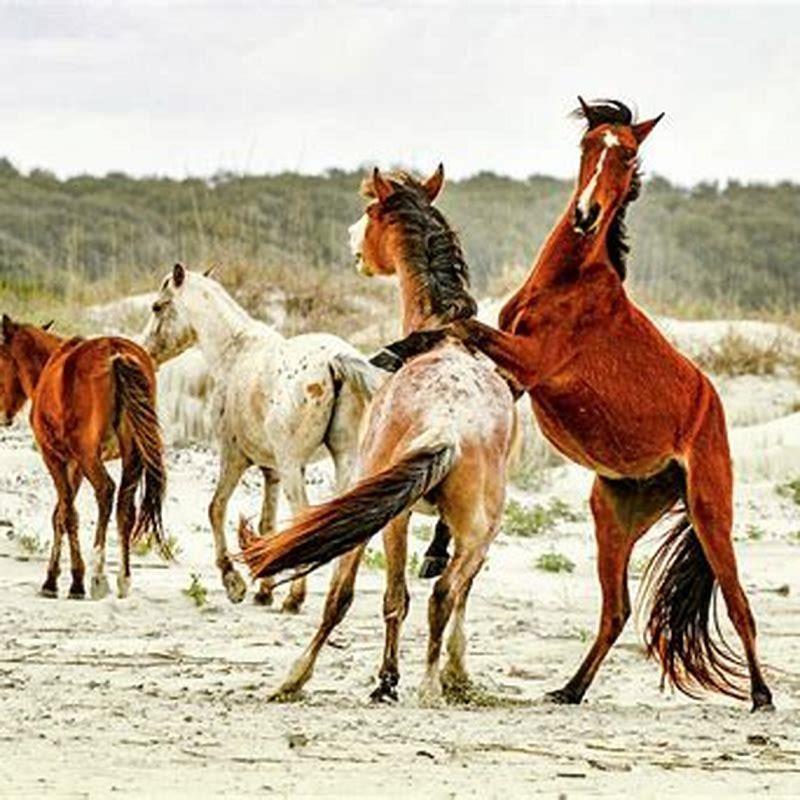
- What does a farrier look for when appraising horse feet?
- What does a farrier use to cut a horse’s feet?
- What does hoof care mean to the horse?
- What happens if a horse does not have a good heel?
- Where does a horse’s hoof begin and end?
- How do you take a farrier Xray of a horse?
- Why is it important to know your horse’s hoof structures?
- What is the function of a horse’s bones?
- When did the Farrier’s Company start?
- Why is a hoof examination so important?
- How do you take a radiograph of a horse?
- Why use radiographs when trimming the equine foot?
- What does an xray show on a horse?
- How do you calibrate a farrier radiograph?
- What does a farrier use a nail file for?
- When did the Farriers become Marshall?
- What did a farrier do in WW1?
- Why is a clinical examination of a horse so important?
- What is the most common radiographic study performed by Equine Practitioners?
- What can a horse radiograph show?
- Where is the X-ray beam centered on a horse?
- Why do radiographs take so long to develop in horses?
- What is an X-ray on a horse?
- What is an equine hoof radiograph?
What does a farrier look for when appraising horse feet?
Farriers develop a fine-tuned sense of smell around horses; if your farrier remarks on the smell of your horse’s feet, pay attention. Using multiple senses to make an appraisal of your horse’s feet makes you less dependent on your eyes and more aware of what is normal for your horse.
What does a farrier use to cut a horse’s feet?
The farrier uses this specialized knife to cut out excess sole and frog in the feet of the horse. Both left-handed and right-handed versions of this knife are available, allowing the farrier to use the appropriate hand depending on the side of the horse he is working on.
What does hoof care mean to the horse?
Given the cost of what this can mean to the horse, one of our primary roles as hoof care providers is to ensure that the horse can land heel-first in its turnout environment and in the riding environment.
What happens if a horse does not have a good heel?
Horses that lack a decent shaped and proper sized heel often suffer from lameness, soreness, and bruising of their hooves due to the lack of protection. The bulbs of the horse’s heel are at the very backmost part of the horse’s hoof.
Where does a horse’s hoof begin and end?
It begins where the bar ends, at mid frog and runs the length of his frog, on the right side. I scrapped the sole next to it so you could see the ridge more clearly. On some horses, it can run around the entire frog, on the sole, from the end of each bar. This hoof created more structure because it is needed.
How do you take a farrier Xray of a horse?
The feet should be thoroughly cleaned, for farrier radiographs the shoes can and should be left in place. The horse should be stood on a flat, level surface. To appreciate bone position, the radiographs should be taken with the horse bearing weight and both feet placed on wooden blocks of equal height.
Why is it important to know your horse’s hoof structures?
“It’s important for horse owners to understand the inner hoof structures for your horse’s soundness and to be able to have meaningful discussions with your farrier,” adds O’Grady. “It helps you open an intelligent conversation based on your experiences, not just something you read online.”
What is the function of a horse’s bones?
Bones (the levers) provide attachment points for a network of ligaments that hold joints together and tendons that connect muscles to bone (the pulleys). Muscles position the bones to make the horse move.
When did the Farrier’s Company start?
It received a Royal Charter of incorporation in 1571. Over the years, the Company has evolved from a trade association for horseshoe makers into an organisation for those devoted to equine welfare, including veterinary surgeons. A farrier’s routine work is primarily hoof trimming and shoeing.
Why is a hoof examination so important?
“A thorough clinical examination is extremely important, of course, which gives invaluable information as to the foot shape, conformation, and quality of the hoof horn, etc. ,” Powell says.
How do you take a radiograph of a horse?
The horse should be stood on a flat, level surface. To appreciate bone position, the radiographs should be taken with the horse bearing weight and both feet placed on wooden blocks of equal height. The cannon bone should be perpendicular to the ground. This can be accomplished by placing a level on the dorsal surface of the cannon bone.
Why use radiographs when trimming the equine foot?
It has evolved to where it quite beneficial for the farrier to use radiographs for guidance when trimming the equine foot. We can immediately see the additional information that can be gained from a radiograph taken of a distorted hoof capsule.
What does an xray show on a horse?
The x-ray will show whether the hoof pastern axis is parallel. If the axis is broken forward (club foot) or if the axis is broken back (long toe underrun heel), the radiograph will reveal the degree of deformity and the best way to trim the foot to improve it.
How do you calibrate a farrier radiograph?
We use a special block with markers of a known distance between them; when we take a radiograph software can automatically calibrate the radiograph and we immediately know distances measured are accurate. The feet should be thoroughly cleaned, for farrier radiographs the shoes can and should be left in place.
What does a farrier use a nail file for?
This is a multi-purpose tool that all farriers use. It is like a nail file for horses and enables the farrier to keep the horse’s hooves even and level if unshod or lightly rasp any hoof that overhangs a shoe.
When did the Farriers become Marshall?
The Ordinances of 1356 were in Norman French, and there is no doubt that in this context ‘Marshalls’ meant ‘Farriers’. Between 1356 and 1674 the Farriers were undoubtedly continuing to practise their craft, and at this time Farriers also treated horses for their ailments.
What did a farrier do in WW1?
The primary job of a farrier was hoof trimming and fitting shoes to Army horses. This combined traditional blacksmith’s skills with some veterinarian knowledge about the physiology and care of horses’ feet. Smiths usually carried the heavy materials they needed with them as they marched.
Why is a clinical examination of a horse so important?
“A thorough clinical examination is extremely important, of course, which gives invaluable information as to the foot shape, conformation, and quality of the hoof horn, etc.,” Powell says.
What is the most common radiographic study performed by Equine Practitioners?
Introduction: Radiographic examination of the digit is the most common radiographic study performed by the equine practitioner. The veterinarian should strive to obtain good quality radiographic equipment and develop good radiographic technique.
What can a horse radiograph show?
• Radiographs can show changes in tissue density, shape, size, outline and position of structures. • Radiographs in the horse are primarily used to assess bones, but can also provide information about soft tissues.
Where is the X-ray beam centered on a horse?
The x-ray beam is centered at the coronary band. Notice in the photo that the cassette is actually placed within a protective holder – this is commonly called a “tunnel.”. This protects the cassette from the weight of the horse. In the photo the square object overlying the coronary band is a lead shield.
Why do radiographs take so long to develop in horses?
• Compared to other species (e.g. humans and dogs) the horse has a very slow bone metabolism, hence radiographic changes are slow to develop. • Once radiographic abnormalities have developed they can persist for a long time without being significant. How does an increase in bone production appear on radiographs?
What is an X-ray on a horse?
An X-ray, or radiograph, is a diagnostic imaging test used to visualize the inner structures of the horse’s body. This procedure involves pointing a machine very similar to a camera at the area of concern, which fires an X-ray beam through the body.
What is an equine hoof radiograph?
Radiology of the equine hoof is used to confirm various disease processes such as laminitis, third phalanx fractures, osteoarthritis (ringbone), navicular disease and extensive hoof wall separations. It has evolved to where it quite beneficial for the farrier to use radiographs for guidance when trimming the equine foot.